Identifying Pigments
- When restoring historical art work, it is important that the new colours used in retouching are a good match of the original colours.
- Chemists can identify the colours and select the most appropriate pigments using a variety of techniques.
Reflectance Spectra
-
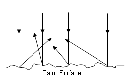
The surface of a painting is never smooth, and as a result light is reflected in all directions.
- A reflectance spectrum gives information on how much light of each colour in the spectrum is reflected in a particular direction:

- The reflectance spectrum of a painting is obtained by shining a light on a small area of the painting and analysing the light reflected off the pigments at a particular angle:

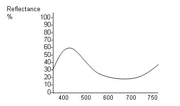
- Once the reflectance spectrums for the different pigment colours are produced, they can be used to select another pigment that is the same colour; for example the reflectance spectrum for a blue-red pigment was measured:

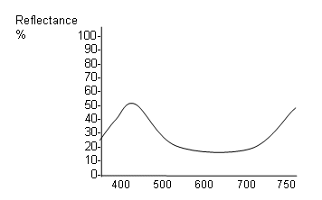
- The reflectance spectrum for ultramarine is as follows:

- These reflectance spectra are very similar and so the colour of the pigments will also be similar. By comparing the spectra of different pigments, the chemists can find the most accurate match of colour.
Laser Microspectral Analysis (LMA)
- This technique is used to identify the different elements present in a pigment.
- A pulse of high power laser light is focused onto a small sample of paint, resulting in the vaporisation of the metal compounds (which have high boiling points).
- A small plume of vapour rises from the paint sample into the region between two electrodes, where the atoms and ions in the vapour are excited to higher electronic energy levels; each element within the compound gives a characteristic emission spectrum that can be used to identify it.
- This method is very sensitive and can be carried out on very small quantities of paint.
- Another technique called energy dispersive x-ray analysis can also be used; this works in a similar way; however the x-ray frequencies are measured rather than the ultraviolet or visible frequencies that are measured in LMA.
Scanning Electron Microscope
- A scanning electron microscope can be used to magnify the pigment by up to 200,000x, allowing the crystal structure of the pigment to be studied.
- Some compounds are different in colour depending on the shape of their crystals; for example lead chromate is darker when the crystals are rod-shaped and lighter when they are flattened.
Useful books for revision
Revise A2 Chemistry for Salters (OCR A Level Chemistry B)
Salters (OCR) Revise A2 Chemistry Home
Home


